Clinical Features

Published on 13/03/2015 by admin

Filed under Neurosurgery

Last modified 13/03/2015

Print this page

rate 1 star rate 2 star rate 3 star rate 4 star rate 5 star
Your rating: none, Average: 0 (0 votes)

This article have been viewed 2552 times

CHAPTER 108 Clinical Features

Neurology of Brain Tumor and Paraneoplastic Disorders

This chapter focuses on three subjects: the pathogenesis of signs and symptoms of brain tumors, the clinical presentation of patients with brain tumors, and the spectrum of central nervous system diseases usually included under the rubric of paraneoplastic neurological disorders. This review is necessarily brief. For a more in-depth treatment of the clinical presentation of brain tumors, see Shapiro and colleagues’ article.1 For the paraneoplastic disorders, see the paper by Darnell and Posner.2

Pathophysiology of Signs and Symptoms Associated with Brain Tumors

Parenchymal brain tumors produce symptoms by three mechanisms: (1) Tumor cell infiltration occurs along nerve fiber tracts in both the white matter and the cortex. Low-grade astrocytomas and oligodendrogliomas behave in this fashion, often producing seizures. (2) Tumor cells grow into a mass, displacing but not destroying the surrounding brain tissue. Metastatic brain tumors typically grow this way. The patient presents with symptoms and signs of an intracranial mass, which often resolve after treatment. (3) A combination of tumor cell infiltration and growth as a mass destroys the surrounding neuropil. Malignant gliomas behave in this fashion, producing symptoms and signs that may not improve after treatment. Although each mechanism takes place independently, considerable overlap occurs, and, as the tumors evolve, all three mechanisms come into play.

Tumors that arise outside of the parenchyma (e.g., meningiomas) rarely infiltrate into the neuropil; instead, they produce their effects by compression of the adjacent brain directly and by the production of cerebral edema. These tumors may present only as a mass, without focal symptoms, or may induce a seizure focus in the underlying brain. As the tumor grows, producing further brain compression, symptoms and signs of brain damage become evident. Resection, especially if carried out when the tumor is small, often restores the patient to a normal neurological state.

Intracranial neoplasms characteristically produce progressive symptoms. The rate of progression varies from an acute apoplectic onset (e.g., after hemorrhage into an intracranial neoplasm, or a seizure associated with cortical stimulation) to progressive mental deterioration. Thus, a patient with a low-grade glioma may have seizures for months to years, developing progressive neurological signs only late in the course of disease. By contrast, a patient with a malignant glioma may develop headaches and focal neurological signs over a few weeks. An acute hemorrhage into a malignant brain tumor may bring the patient to the hospital abruptly with little warning that an intracranial tumor has been developing. Similarly, it is not unusual for a patient to seek psychiatric help because of depression or change in work habits, only to be found to harbor a cerebral neoplasm. It is the progressive nature of the symptoms that alerts the clinician to suspect brain tumor.

Brain tumors produce generalized symptoms because of their expanding mass effect. They produce focal symptoms by direct compression on, or infiltration into, the surrounding neuropil.

Generalized symptoms from brain tumors are manifestations of increased intracranial pressure, a result of the expanding tumor volume and the associated edema. Tumors may obstruct the cerebrospinal fluid (CSF) pathways, producing hydrocephalus. As the mass enlarges, intracranial pressure rises; brain tissue may be displaced through the rigid intracranial dural passageways, producing various herniation syndromes. Cerebral dysfunction and headache ensues. Abrupt headache and worsening neurological signs may accompany sudden increases in intracranial pressure that last 5 to 20 minutes. These episodes of raised pressure have a characteristic appearance on recording of intracranial pressure leading to their name, plateau waves.3

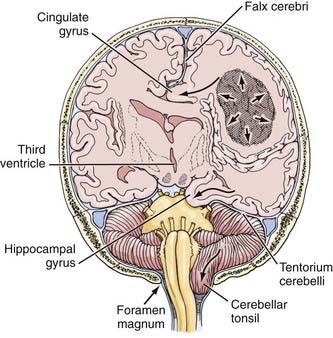
The herniation syndromes are life-threatening. Because the skull is divided into compartments by the relatively inelastic dura, an expanding mass in one compartment forces brain tissue through the openings between compartments, tearing blood vessels and compressing the neuropil (Fig. 108-1).4 A unilateral cerebral mass may force the medial surface of the brain beneath the falx cerebri (cingulate herniation). Transtentorial and tonsillar herniation may occur because of displacement of brain tissue through, respectively, the tentorial notch and foramen magnum. Table 108-1 lists the pathogenesis and clinical manifestations of the herniation syndromes.4

TABLE 108-1 Herniation Syndromes

PATHOGENESIS CLINICAL MANIFESTATION
CENTRAL HERNIATION
Lateral and Forward Shift of Diencephalon and Upper Midbrain
Diencephalic compression Decreased state of consciousness, small pupils, Cheyne-Stokes respiration
Midbrain pontine compression

Lower pons-medullary compression Tonsillar (Foramen Magnum) Herniation Posterior fossa mass pushes cerebellar tonsils through foramen magnum, compressing medulla Rapid loss of consciousness, stiff neck, vomiting, skew deviation of eyes, irregular respirations, death UNCAL HERNIATION Temporal Lobe Herniation through Tentorial Notch, Compressing Ipsilateral third nerve Third nerve palsy Posterior cerebral artery Homonymous hemianopia Midbrain   Opposite cerebral peduncle Ipsilateral hemiparesis

Adapted from Posner JB, Saper CB, Schiff N, Plum F. Plum and Posner’s Diagnosis of Stupor and Coma, 4th ed. New York: Oxford University Press, 2007.

The clinical manifestations of transtentorial herniation depend in part on the site of the mass lesion. Central herniation occurs when the mass effect is bilateral or becomes symmetrical, causing a downward shift of the diencephalon and upper brainstem. Uncal herniation occurs when the mass is unilateral, especially temporal, and the medial temporal lobe is displaced inferiorly and medially through the tentorial notch.

The clinical manifestations of the two herniation syndromes overlap, although there are differences in the specific nature and temporal sequence of individual symptoms. With central herniation, diencephalic compression produces decreased state of consciousness, small pupils, and respiratory abnormalities. With progression, the pupils become midposition and fixed, respirations are frequently Cheyne-Stokes; decorticate and decerebrate posturing follows; and progressive coma ensues. Ultimately, apnea, slow pulse, and raised blood pressure presage death. In uncal herniation, the ipsilateral third nerve may be compressed, leading to partial oculomotor palsy, with paresis of extraocular movement and a dilated pupil. The posterior cerebral artery may be compressed against the tentorium, producing a homonymous hemianopia. Further progression is identical to that of central herniation. Occasionally, the opposite cerebral peduncle may be compressed against the tentorium, producing an ipsilateral hemiparesis, one of the false localizing signs (i.e. neurological signs that incorrectly suggest a location of a specific lesion). They also include unilateral or bilateral lateral rectus palsy from sixth nerve compression. False localizing signs are more common in slowly growing neoplasms like meningiomas.

Although tonsillar herniation can be caused by supratentorial mass lesions, a more common cause is an expanding posterior fossa mass, which forces the cerebellar tonsils through the foramen magnum, compressing the medulla. The patient becomes obtunded, with headache, vomiting, stiff neck, and, occasionally, opisthotonos. Other signs include skew deviation of the eyes and other disconjugate eye movements, arterial hypertension, and syncope with cough or sudden postural change. Respiratory activity is irregular, and acute apnea may occur. Upward herniation may occur with rapidly expanding cerebellar masses, wherein the brainstem is forced upward through the tentorial notch. Lumbar puncture may produce tonsillar herniation acutely.

Cerebral edema accompanies growing brain tumors. It is produced mostly within a parenchymal brain tumor but also comes from the surrounding brain tissue as a result of the release of vasogenic permeability factors produced by the tumor, which act on brain capillaries.5 (The vascular permeability factor has been identified as vascular endothelial growth factor, which plays an important role in angiogenesis of tumors.6) Cerebral edema increases the total size of the mass. It is not unusual for a tumor of perhaps 20 g to produce a mass of nearly 100 mL because of the associated edema. Cerebral edema may also be produced by an extra-axial tumor, in which case the edema, also vasogenic in origin, comes only from the brain tissue itself.

Treatment of cerebral or cerebellar herniation includes corticosteroid hormones (e.g., dexamethasone, 10 mg intravenously, followed by oral divided doses of 16 mg/day), intravenous mannitol 25 to 50 g, and sometimes intubation and hyperventilation to reduce plasma carbon dioxide and thus reduce cerebral blood flow. Subsequent treatment depends on the site and nature of the tumor.

Focal symptoms occur as a direct effect of the tumor’s growth into the surrounding neuropil. As the tumor infiltrates along nerve fiber tracts or surrounds neurons (satellitosis), it ultimately interferes with neurological function. “Irritation” from the tumor on adjacent cortex may produce seizures. Seizures are a common manifestation of intracranial tumor, both generalized and focal. More commonly, the tumor produces loss of neurological function, manifested as weakness or sensory disturbances. Finally, the tumor itself may hemorrhage, producing acute headache and focal localizing signs. Tumors that are particularly likely to hemorrhage include glioblastomas and metastatic tumors from melanoma, choriocarcinoma, and testicular carcinomas.

Clinical Manifestations of Intracranial Tumors

The symptoms and signs of intracranial tumor depend on the size of the tumor, its location, and its rate of growth. Manifestations may be general or focal, or both. General manifestations include mental changes, headache, generalized convulsions, nausea, and vomiting. Examples of focal signs include focal seizures, weakness, sensory abnormalities, speech disturbances, and visual defects. It is important to recognize that general and focal manifestations overlap, as does their pathogenesis. Thus, although mental dullness often accompanies large masses, mental changes may occur with small focal tumors in the temporal lobe, or may be part of an aphasia syndrome. In addition to its size, the location of the tumor in the brain quantitatively influences signs and symptoms. Thus, a small tumor in the speech or visual association cortex may produce more signs than does a large frontal tumor. In addition, the character of an abnormality may differ according to whether it accompanies a generalized increase in intracranial pressure or is part of a specific focal dysfunction.

General Manifestations

Mental Changes

Mental changes are a frequent general clinical manifestation of intracranial tumor (Table 108-2). They are often subtle in presentation and onset and may not attract the attention of friends and family members until the patient’s behavior becomes abnormal. Psychomotor retardation is the most common mental change accompanying a brain tumor. Characteristically, impersistence in routine tasks, emotional lability, inertia, faulty insight and forgetfulness, reduction in the range of mental activity, indifference to social practices, reduced initiative and spontaneity, and blunted affect are seen. Patients may sleep for longer periods, complaining that they seem unable to get through a day without taking a nap. Frank confusional states and dementia usually occur later and are more likely to accompany focal signs of brain disease. Alternatively, the change in the state of consciousness may progress to stupor and frank coma. Changes in personality may be described in “psychological terms”: depression, euphoria, impulsive behavior.7 In my series, almost 20% of patients have, at one time or another, sought psychological help for symptoms that, in retrospect, were clearly related to the tumor. Mental changes must be viewed together with other signs and symptoms, and especially with a history of progression of symptoms, to lead the physician to suspect a neoplastic process.

TABLE 108-2 Mental Changes Accompanying Brain Tumors

PSYCSHOMOTOR RETARDATION

SLEEP DISTURBANCES

LANGUAGE DISORDERS

SOCIAL DISTURBANCES “PSYCHOLOGICAL” SYMPTOMS

Headache

Headache occurs from traction of basal meningeal structures and is worse the larger the mass, although even small tumors may produce such traction, especially meningiomas. The degree and intensity of headache also relate to the rapidity with which traction develops. A rapidly enlarging intracranial mass may produce sudden severe headache, whereas a slowly growing mass may become quite large without producing headache.

Headache occurs in about 50% of patients with brain tumor at some time during the course of the illness.8 Headache is more prominent and tends to occur earlier when the tumor produces increased intracranial pressure. Of special importance is a headache that has recently changed in character, for example, becoming more intense, frequent, or longer lasting. Although classic brain tumor–associated headaches are severe, often worse in the morning, all brain tumor patients do not have morning headaches, and in fact may have head pain at any time of day or night.8 The head pain from brain tumor is often intermittent and may be described as deep, aching, or pressure-like rather than the more characteristic throbbing pain of migraine. It is often aggravated by the Valsalva maneuver, as in coughing or straining, and frequently becomes worse through change of posture. The headache that occurs in the early morning hours appears to be related to the recumbent posture and is thought to be associated with increased intracranial pressure as a result of lying down.

Generalized Convulsions

Seizures occurring for the first time in adults are more likely to be due to focal cerebral disease, especially neoplasms, than are seizures that begin in childhood, which are more often part of the epilepsies that are characteristic of this age group.9 Intracranial tumors produce both generalized major motor convulsions and various forms of focal seizures. Petit mal epilepsy is probably never due to a neoplasm, but minor temporal lobe seizures (partial simple or complex) that resemble petit mal attacks may accompany temporal lobe tumors. Jacksonian seizures that progress from one body part to another usually imply a lesion of the motor or sensory cortex. Lesions of the temporal lobe characteristically give rise to partial complex (psychomotor) seizures that may be associated with olfactory hallucinations (uncinate fits), disorders of visual or auditory perception, episodes of déjà vu, or automatic behavior.

Seizures, both generalized and focal, occur in 25% to 50% of patients with cerebral tumors (Table 108-3).10 They are more likely to accompany slowly growing tumors than they are the more rapidly growing malignant neoplasms.11 A review of data of the Brain Tumor Cooperative Group (BTCG) database revealed that seizures occurred as the initial manifestation of tumor in 50% of patients with anaplastic astrocytoma but in only 27% of patients with glioblastoma multiforme. Seizures have been reported to occur in 25% of patients with reported malignant gliomas.12

Buy Membership for Neurosurgery Category to continue reading. Learn more here