108 Clinical Assessment of Renal Function
Five to 15 percent of patients in intensive care units (ICUs) experience acute deterioration in renal function.1,2 Conversely, renal dysfunction adds substantially to the morbidity and mortality of these patients. Moreover, changes in renal function directly affect drug disposition. Thus, a means to assess renal function is essential for optimal management. The glomerular filtration rate (GFR) is the standard measure of renal function. It reflects overall renal functional capacity and, in renal failure, correlates with structural damage to the kidney. This chapter reviews selected aspects of renal physiology with an emphasis on measurement of renal function, consequences of altered function, and approaches to improving renal function. The focus is on measurement and optimization of glomerular filtration rate (GFR) and renal blood flow (RBF).
 Renal Blood Flow
Under physiologic conditions, blood flow to the kidneys is 20% of cardiac output. This high rate of blood flow (1-1.2 L/min) is particularly remarkable in that the kidneys make up only 0.5% of total body weight. The high blood flow rate is due, at least in part, to the unique anatomic arrangement of the renal vasculature, with the interlobar and arcuate vessels offering little resistance to flow. This in turn is because the interlobular arteries originate from the arcuates in a parallel arrangement and because the afferent arterioles also arise in a parallel arrangement from the interlobular vessels. It is this parallel arrangement that accounts for the low resistance, because the total resistance of n equals parallel paths, each with a resistance R, is R/n3. Major resistance vessels in the kidney are the afferent and efferent arterioles that bound the glomerular capillary network. Although total resistance is a function of resistance across each of these vessels, it is a unique feature of the kidney that variations in the individual resistances across the afferent and efferent arterioles, respectively, may lead to alterations in glomerular capillary pressure and, hence, in GFR.3
Despite a wide range of perfusion pressures, RBF and GFR are maintained relatively constant, a process described as autoregulation. The term autoregulation generally refers to the relative constancy of GFR over a range of perfusion pressures but also refers to the regulation of RBF. Emphasis has been placed on the preglomerular vasculature, mainly the afferent arterioles, as the major site at which renal perfusion is regulated. However, studies also suggest that the larger vessels, such as the interlobular vessels, may respond to a variety of vasoactive stimuli and participate in an autoregulatory phenomenon. A variety of hypotheses have been generated to explain the autoregulatory response of the kidney with respect to RBF. There is evidence to suggest mediation by neural, humoral, or intrarenal factors that regulate the renal circulation.4
Other vasoactive compounds that affect the renal circulation include the plasma and glandular kallikreins and kinins and endothelium-derived vasoactive factors such as nitric oxide and endothelin.4 Among the catecholamines, α- and β-adrenergic agonists are known to affect renal vascular tone by causing vasoconstriction and vasodilatation, respectively. In addition, dopamine in low doses leads to renal vasodilatation. Emphasis has more recently been placed on atrial natriuretic peptide and purinergic agents such as adenosine. The effect is likely to be influenced by changes in salt intake and extracellular fluid volume as well as by hydration status. For example, the influence of AII on renal hemodynamics is greater in sodium depletion, which also activates the sympathetic nervous system. In response to mild nonhypotensive hemorrhage, renal hemodynamics are relatively well maintained. However, with further reductions in volume associated with a more severe hemorrhage, renal ischemia mediated by activation of the renin-angiotensin system, renal efferent adrenergic nerves, and circulating catecholamines may occur.4
Finally, modification of dietary protein and amino acid intake may affect renal hemodynamics. Dietary protein intake in excess of 1 g/kg/d has been associated with renal vasodilatation, as have infusions of casein hydrolysates and amino acids.5,6 Conversely, chronic consumption of a low-protein diet may be associated with renal vasoconstriction.
Measurement of Renal Blood Flow
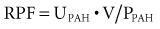
where UPAH and PPAH refer to urine and plasma PAH concentration, respectively, and V is urine flow rate in milliliters per minute.
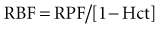
RBF can be estimated by correction for the hematocrit (Hct):
Although available, this test is rarely used in clinical practice. In fact, direct quantitation of RPF and RBF is rarely indicated outside research studies; however, sometimes it is necessary to document that the kidneys are being perfused. In this case, one of three additional methods may be utilized: (1) selective arteriography, including CT angiography and MR angiography, (2) Doppler ultrasonography, and (3) external radionuclide scanning.
Because the latter two methods are noninvasive, they are preferred. With respect to the nuclide study, until recently, scanning was usually performed utilizing 125I-iodohippurate sodium; however, the poor radiologic characteristics of 131I limit its use in renal imaging.7 More recently, other agents such as 127I-orthoiodohippurate and 99mTc-L, L-ethylenedicysteine may prove to be superior.7,8
Clinical Correlates
Although a significant body of data has been obtained to indicate a complex relationship between neurocirculatory factors and renal hemodynamics, several points can be made from a clinical perspective. Optimization of cardiac output and extracellular fluid (ECF) volume, including the intravascular space, is essential for the maintenance of renal perfusion. Particularly because the effects of vasoactive compounds such as AII and catecholamines are accentuated in the presence of renal hypoperfusion and volume contraction, attention should be directed to an assessment of ECF volume, with correction of any deficits, and to optimizing cardiac function. Frequently, pharmacologic agents have been employed to maintain renal perfusion in situations in which this may be compromised. Specifically, there has been widespread use of so-called low-dose or renal-dose dopamine infusions. This is based on the observation that in low doses (<3 µg/kg/min) dopamine leads to renal vasodilatation.9 At higher doses, renal vasoconstriction may occur.
The beneficial effects of dopamine infusion have not been documented in patients who are depleted of sodium chloride and volume, and the use of dopamine has not been shown to be effective beyond a short period of infusion.9–11 That is, infusions of renal-dose dopamine for 24 to 36 hours may be beneficial in the appropriate circumstance, but there is no evidence supporting the long-term use of this agent. Thus, justification for prolongation of its use beyond several days is not supported by available data. Furthermore, reports suggest that adverse outcomes may be associated with the use of dopamine.11 Continuous infusions of fenoldopam mesylate, a potent dopamine A-1 receptor agonist, have been employed in an attempt to preserve renal function in a variety of clinical settings. A meta-analysis of 16 randomized trials in critically ill patients showed that fenoldopam significantly reduced the risk of acute kidney injury, need for renal replacement therapy, and in-hospital death.12 Beyond anecdotal evidence, there are no compelling data to support the use of other potential vasodilator substances such as prostaglandins. Although high-protein feeding and amino acid infusions may increase RBF by an undefined mechanism, there is no justification for utilizing these therapies solely from a hemodynamic point of view.5,6
 Glomerular Filtration Rate
Measurement of Glomerular Filtration Rate
More recently, other radiolabeled nuclides have been found to be satisfactory substitutes for inulin and have advantages in the measurement of GFR.7,8,13,14 Particularly 99mTc-labeled diethylenetriamine pentaacetic acid (DTPA) and 125I- or 131I-labeled iothalamate clearances closely approximate the CIn.15,16 99mTc-DTPA has been utilized and found to give measurements that correlate closely with CIn in ICU patients.17,18 In addition, the clearance of gentamicin has been utilized in a limited fashion to measure GFR.19,20 At the present time it is not common for GFR to be measured directly. Rather, GFR is estimated by the endogenous creatinine clearance or serum creatinine determination (see later).
The normal values for GFR given previously apply for individuals from the teenage years through approximately age 35. Thereafter, GFR declines in most individuals. Whereas this decline was formerly thought to occur at a relatively constant rate of approximately 10 mL/min per decade,21–23 more recent data obtained in a longitudinal fashion indicate that this reduction is not so predictable.24 In addition, a circadian rhythm for GFR has been described.25,26 GFR is maximal in the daytime, whereas a minimal value during the night has been found in normal individuals. Whether this circadian pattern of GFR occurs in critically ill hospitalized patients is not known.
 Creatinine Clearance and Serum Creatinine
Creatinine Clearance
The endogenous creatinine clearance (CCr) enjoys widespread use as a reasonable gauge of GFR when great precision is not demanded, which it rarely is in clinical practice. The use of creatinine as a marker of GFR has the advantage that creatinine is endogenously produced and is easily measured by inexpensive methods. Creatinine, like inulin, is freely filtered and absorbed minimally if at all by the tubules. However, creatinine is secreted, and the contribution of secretion to total excretion is greater as the GFR decreases and serum creatinine rises. At GFRs below 40 mL/min, CCr exceeds CIn by 50% to 100%.15,27 When GFR is significantly depressed and it is deemed important to get a more precise measurement of GFR, one of the previously mentioned methods to estimate GFR directly might be utilized. Additionally, because CCr overestimates GFR and the clearance of urea underestimates GFR, the mean value of simultaneously obtained creatinine and urea clearances has been shown to provide a close estimation of CIn when the latter is below 20 mL/min.28
Because cimetidine competes with creatinine for tubular secretion (see later), administration of cimetidine may increase the accuracy both of creatinine clearance in 24-hour collections (when given for several days beforehand) and of 4-hour, water-loaded clearances.29–31 Taking advantage of this effect results in a more accurate estimate of GFR. Specifically, CCr obtained in the presence of cimetidine (400 mg as a priming dose followed by 200 mg every 3 hours) yielded values that closely approximated CIn.29,30 Volume expansion in humans causes a small rise in GFR, whereas volume depletion, severe heart failure, hypotension, anesthesia, surgery, trauma, sepsis, and even mild intestinal bleeding without frank hypotension may depress GFR substantially.
Various methods are available to measure creatinine. Creatinine is frequently measured using the Jaffé alkaline picric acid reaction. Although this method is widely utilized, this reaction also measures other chromogens, which may lead to a false elevation in the estimated serum creatinine (SCr) measurement. Substances such as acetoacetate (in ketoacidosis), pyruvate, ascorbate, 5-flucytosine, certain (but not all) cephalosporin antibiotics, and very high urate artifactually raise SCr in normal subjects by 0.5 to 2 mg/dL.32–38 These substances are excreted into the urine but contribute trivially compared with overall urine creatinine (UCr). Thus, noncreatinine chromogens affect the SCr but have little effect on the UCr.
In individuals with normal renal function, the contribution of serum noncreatinine chromogens to raising the SCr is approximately equal to the contribution of secretion to creatinine excretion, such that the CCr closely approximates GFR. As GFR decreases, the contribution of noncreatinine chromogens to the total measured SCr becomes less than the secreted moiety, and the CCr overestimates GFR to a greater extent. Direct enzymatic creatinine measurements are not affected by noncreatinine chromogens. Very high levels of serum glucose (>1000 mg/dL) and 5-flucytosine may interfere with the enzymatic reaction, whereas high levels of bilirubin (>5 mg/dL) affect the autoanalyzer method36 and lead to falsely low SCr values. It is therefore important to know the method by which a given laboratory measures SCr. Competing for the same proximal tubular organic base secretory site as creatinine, certain pharmacologic agents may suppress this process and lead to a rise in SCr. Trimethoprim, probenecid, and cimetidine, but not ranitidine, are organic bases that inhibit creatinine secretion competitively and can result in a mild elevation in SCr, usually 0.5 mg/dL or less.39–42
As with all clearance methods, the CCr is subject to errors that may amount to as much as 10% to 15% or more. In addition to potential problems in estimating SCr and UCr, errors in timing of urine collection, incomplete collection, and inaccurate measurement of urine volume are other factors that contribute to errors.43 Although 24-hour UCr clearances have been widely utilized, no specified time period is required for the clearance to be obtained. In fact, shorter collection periods of several hours may be more accurate in patients passing adequate amounts of urine (not oliguric), particularly if the patient is not in a steady state (see later). To reduce errors in volume measurement, one can induce a water diuresis in stable subjects before beginning the test,44
