Choledochal Cyst and Gallbladder Disease
Choledochal Cyst
Choledochal cyst is a congenital dilatation of the biliary tract. The dilatation can be found along any portion of the biliary tract. However, the most common site is the choledochus. The diameter of the bile duct varies according to the child’s age.1–3 Normal diameter of the common bile duct (CBD) is seen in Table 44-1.2 Any diameter of the bile duct greater than the upper limit should be considered abnormal.
TABLE 44-1
The Mean Common Bile Duct Diameter and the Range According to a Patient’s Age
| Age (Years) | Range (mm) | Mean (mm) |
| ≤4 | 2–4 | 2.6 |
| 4–6 | 2–4 | 3.2 |
| 6–8 | 2–6 | 3.8 |
| 8–10 | 2–6 | 3.9 |
| 10–12 | 3–6 | 4.0 |
| 12–14 | 3–7 | 4.9 |
Adapted from Witcombe JB, Cremin BJ. The width of the common bile duct in childhood. Pediatr Radiol 1978;7:147–9.
Classification
Different classifications have been proposed for choledochal cyst.4–8 However Todani’s classification has been the most widely accepted (Fig. 44-1).5 According to this classification, choledochal cyst (CC) are classified into five types:
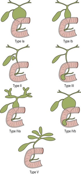
FIGURE 44-1 These diagrams depict the five classifications for choledochal cyst according to Todani. (From Todani T, Watanabe Y, Narusue M, et al. Congenital bile duct cyst: Classification, operative procedure, and review of 37 cases including cancer arising from choledochal cyst. Am J Surg 1977;134:263–9.)
• Type II: diverticulum of the CBD
• Type III: choledochocele (dilatation of the terminal CBD within the duodenal wall)
• Type V—intrahepatic duct cyst (single or multiple, as in Caroli disease).
Forme fruste is a special variant of choledochal cyst characterized by pancreaticobiliary malunion, but little or no dilatation of the extrahepatic bile duct.8–12 Children present with symptoms similar to those in patients with a CC. Excision of the extrahepatic bile duct is recommended in children because of the likely eventual development of cancer due to chronic pancreaticobiliary reflux.
Type I CCs predominate. Together with type IVa cysts, they account for more than 90% of cases. Caroli disease is characterized by segmental saccular dilatation of the intrahepatic bile ducts. It may affect the liver diffusely or be localized to one lobe.13–15
Etiology
There are many theories to explain the development of a CC. However, none of these can explain the formation of the five different types of CC. Choledochal cysts seem to be either congenital or acquired. Congenital cysts develop during fetal life.16 These appear to develop as a result of a prenatal structural defect in the bile duct. Shimotake found that the total number of ganglion cells within the wall of these CCs is significantly lower than in control specimens.17 Also, smooth muscle fibers are more abundant in the cystic type than in the fusiform type.18
Choledochal cysts which develop later in life are considered ‘acquired’.16 The theory of the long common biliopancreatic channel proposed by Babit has been widely accepted to explain the formation of this type.19 Normally, the terminal CBD and pancreatic duct unite to form a short common channel, which is well surrounded by Oddi’s sphincter. This normal anatomic arrangement prevents the reflux of pancreatic fluid into the bile duct. If this common channel is long and part of it is not surrounded by the normal sphincter, pancreatic secretions can reflux into the biliary tree (Fig. 44-2). Proteolytic enzymes from the pancreatic fluid are activated and can cause epithelial and mural damage that leads to mural weakness and dilatation of the choledochus. This theory is supported by the fact that high concentrations of activated pancreatic amylase and/or lipase have been found in patients with CC and long pancreaticobiliary channels.16,20–22 In an experimental study, CC was produced by creating a choledochopancreatic end-to-side ductal anastomosis.23 Also, a high incidence of a common channel has been detected in CC patients.24
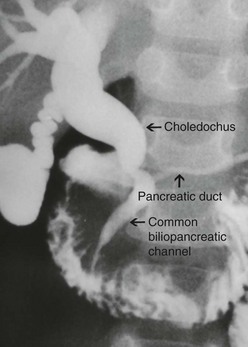
FIGURE 44-2 This contrast study depicts a long common biliopancreatic channel which allows reflux of pancreatic secretions into the biliary tree. A long common biliopancreatic channel is thought to be the etiology of an acquired choledochal cyst.
Obstruction at the level of the pancreaticobiliary junction may be an associated causal factor in choledochal dilatation. An experimental model for the study of cystic dilatation of the extrahepatic biliary system has been produced by ligation of the distal end of the CBD in the newborn lamb.25 High biliary pressure in patients with CC have also been recorded during operative correction.26
In adults, an anomalous union of the pancreaticobiliary duct is defined when the common pancreaticobiliary channel is longer than 15 mm,3,27 or when its extraduodenal portion is more than 6 mm.28 In a study of 264 infants and children undergoing endoscopic retrograde cholangiopancreatography (ERCP), the maximal length of the common channel was found to be 2–7 mm among children ≤ 3 years old, 4 mm among children 4 to 9 years old, and 5 mm between 10 and 15 years old.29
Clinical Features
Females are affected more often than males. In our series of 400 cases, the female to male ratio was 3.2 to 1.30 Clinical presentations differ according to the age of onset and the type of cyst. An abdominal mass or jaundice is a common finding in an infant with CC, whereas abdominal pain is more often seen in older children.16,31–33 The cystic form usually presents with an abdominal mass whereas the fusiform type is usually found in patients presenting with abdominal pain. Choledochal cysts diagnosed antenatally are more likely cystic in nature.16
Clinical manifestations among our 400 cases included: abdominal pain (88%), vomiting (46%), fever (28%), icterus (25%), discolored stool (12%), abdominal tumor (7%), and the classic triad (2%).30
Complications such as perforation and hemobilia are rare;34,35 however pancreatitis is common.16,31 Malignant change is a late complication, mostly seen in adults.36–39
Imaging
Ultrasonography (US) is the initial imaging method of choice (Fig. 44-3A). Contour and position of the cyst, the status of the proximal ducts, vascular anatomy, and hepatic echotexture can all be evaluated on ultrasound.
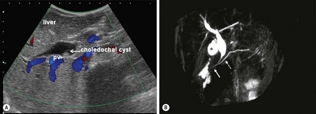
FIGURE 44-3 (A) Ultrasound is the initial imaging method of choice for identifying a choledochal cyst. The cyst is identified as well as the portal vein (PV) lying posterior to it. (B) MRCP is highly accurate in the detection and classification of the cyst. On this MRCP image, note the fusiform choledochal cyst as well as the pancreatic duct (dotted arrow) and long common channel (solid arrow). The gallbladder is marked with an asterisk.
ERCP allows excellent definition of the cyst as well as the entire anatomy, including the pancreatobiliary junction. However, this investigation is invasive and has complications such as pancreatitis, perforation of the duodenal or biliary tracts, hemorrhage, and sepsis.40,41
Magnetic resonance cholangiopancreatography (MRCP) is highly accurate in the detection and classification of the cysts. (Fig. 44-3B) The overall detection rate of MRCP for a CC is very high (96–100%) and should be considered a first-choice imaging technique for evaluation.42–44
Intraoperative cholangiography is indicated when the anatomic detail of the biliary tract cannot be demonstrated by MRCP or ERCP (Fig. 44-4). Contrast-enhanced CT may be indicated in some patients with pancreatitis or if an associated tumor is suspected.
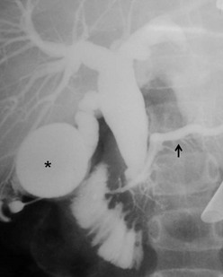
FIGURE 44-4 In this patient, neither an MRCP nor ERCP were helpful preoperatively. Thus, an intraoperative cholangiogram was performed to identify the anatomic detail within the biliary tract. The distended gallbladder is marked with an asterisk. The enlarged choledochal cyst is seen and the pancreatic duct is identified with a solid arrow.
Surgical Techniques
General Principles
Cystoduodenostomy and cystojejunostomy have been abandoned due to cholangitis, stone formation, and malignant degeneration.6,45–47 External drainage is indicated for a perforated cyst in patients whose condition is too unstable to perform cystectomy and a bilio-enteric anastomosis.34,35
Cyst excision and a bilio-enteric anastomosis is the preferred approach for most patients. The cyst should be excised at the level of the common biliopancreatic channel orifice at its distal end and approximately 5 mm from the confluence of the right and left hepatic ducts at the proximal end. Postoperative malignancy in a residual cyst on either hepatic duct side or from the distal part has been reported.48,49 A review from the English language and Japanese literature of 23 patients with carcinomas of the bile duct developing after CC excision found that malignancy developed in the intrapancreatic remnant of the bile duct or CC in six patients, in the remnant of the CC at the hepatic side in three patients, in the hepatic duct at or near the anastomosis in eight patients, and in the intrahepatic duct in six patients.48 Abdominal pain and pancreatitis due to leaving a remnant of the cyst in the pancreatic head has also been described.50
Operative correction can be performed safely in all age groups.30,51–53
Bilio-Enteric Anastomosis after Cystectomy
Many surgeons use hepaticojejunostomy54–59 while others prefer hepaticoduodenostomy.60–64 Fat malabsoption and duodenal ulcer are the main concerns with a hepaticojejunostomy. In addition, the operative time is longer in comparison with hepaticoduodenostomy. Complications after Roux-en-Y hepaticojejunostomy such as a twist of the Roux limb, intestinal obstruction, and duodenal ulcers have been reported.65–68 On the other hand, cholangitis and gastritis due to bilious reflux are the main concerns with hepaticoduodenostomy.69,70 However, the operative time is shorter in comparison with hepaticojejunostomy. A hepaticoduodenostomy is considered more physiologic because the bile drains directly into the duodenum. This anastomosis is performed above the transverse colon mesentery which may help prevent intestinal obstruction from adhesions.
Laparoscopic Approach
Endotracheal intubation general anesthesia is standard. Epidural analgesia can provide excellent postoperative pain relief. Broad spectrum intravenous antibiotics are best given at induction of anesthesia and continued for five days postoperatively. A nasogastric tube, rectal tube, and urinary catheter are used to decompress the stomach, colon, and bladder. The patient is placed in a 30° lithotomy position (Fig. 44-5). The surgeon stands at the lower end of the operating table between the patient’s legs.
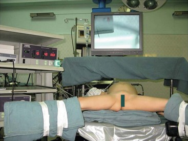
FIGURE 44-5 Older patients are placed in the lithotomy position and smaller patients are moved to the end of the bed. It is helpful for the surgeon to stand either between the patient’s legs (in older patients) or at the end of the operating table (in younger patients) for laparoscopic choledochal cyst repair.
The first laparoscopic operation for CC was reported in 1995.71 This approach quickly became popular and has become a routine procedure in many centers.30,72–84 The laparoscopic approach is preferred for most types of CC: I, II, and IV. Relative contraindications are in patients with perforation, previous biliohepatic surgery, or especially newborns with damaged hepatic functions.
A 10 mm cannula is inserted through the umbilicus for the telescope. Three additional 5 or 3 mm ports are introduced for the working instruments: one in the right flank, another in the left flank, and one in the left hypochondrium (Fig. 44-6). Carbon dioxide pneumoperitoneum is maintained at a pressure of 8–12 mmHg. The liver is secured to the abdominal wall with a suture placed around the round ligament (Fig. 44-7A). The cystic artery and the cystic duct are identified, clipped, and divided. A second traction suture is placed at the junction of distal cystic duct and gallbladder fundus to elevate the liver and expose the hepatic hilum (Fig. 44-7B).
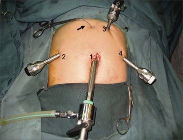
FIGURE 44-6 This operative photograph depicts placement of the ports for laparoscopic repair of a choledochal cyst. A 10 mm cannula (1) is introduced through the umbilicus for the telescope. Three additional 5 mm or 3 mm ports are then used for the working instruments (2,3,4). Note the liver has been elevated anteriorly with a suture placed around the round ligament and exteriorized in the epigastric region (arrow).
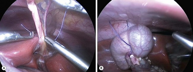
FIGURE 44-7 (A) Suture has been placed through the round ligament and will be exteriorized in the epigastrium in order to help elevate the liver for exposure of the choledochal cyst. (B) A second traction suture has been positioned at the junction of the distal cystic duct and gallbladder fundus to further elevate the liver anteriorly and expose the hepatic hilum.
The inferior part of the cyst is separated from the pancreatic tissue down to the common biliopancreatic duct using a 3 mm dissector for cautery and dissection. Protein plugs or calculi within the cyst and common channel are washed out and removed. The inferior part of the cyst is opened longitudinally and inspected to identify the orifice of the common biliopancreatic channel. A small catheter is then inserted into the common channel. Irrigation with normal saline via this catheter is performed to eliminate protein plugs until clear fluid returns and the catheter can be passed down into the duodenum (Fig. 44-8A). A cystoscope can be used to remove protein plugs in the common channel if its diameter permits.85,86 The distal CC is then clipped and divided at the level of the orifice of the common channel (Fig. 44-8B).
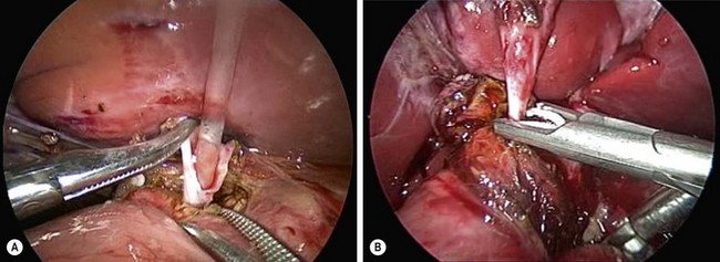
FIGURE 44-8 (A) After opening the inferior part of the cyst to identify the orifice of the common biliopancreatic channel, a small catheter is inserted into the common channel for irrigation and elimination of protein plugs. (B) After irrigating the common channel, the distal choledochal cyst is being ligated with an endoscopic clip and will subsequently be divided at the level of the orifice of the common channel.
The cephalad portion of the cyst is further dissected to the common hepatic duct. The cyst is initially divided at the level of the cystic duct. After identifying the orifice of the right and left hepatic ducts, the rest of the cyst is removed, leaving a stump approximately 5 mm from the bifurcation of the hepatic ducts. Irrigation with normal saline through a small catheter inserted into the right and then into the left hepatic duct is performed to wash out protein plugs or calculi until the effluent is clear.
Hepaticojejunostomy
The Roux limb is passed through a window in the transverse mesocolon to the porta hepatis. The jejunum is opened longitudinally on its antimesenteric border a few millimeters from the end of the Roux limb to avoid the creation of a significant blind pouch as the child grows. The hepaticojejunostomy is fashioned using two running sutures of 5-0 PDS (interrupted sutures are used when the diameter of the common hepatic duct is less than 1 cm). The anastomosis is performed from left to right using 3 mm instruments. If the diameter of the common hepatic duct is too small, ductoplasty is performed by opening the common hepatic duct and incising the left hepatic duct longitudinally for a variable distance.
Alternative Operative Techniques
An alternative biliary reconstruction technique is an end-to-end hepaticojejunostomy (in contrast to end-to side). This should only be performed if there is no significant size discrepancy between the hepatic duct and Roux limb. A hepaticoduodensotomy with jejunal interposition can be performed if desired. The robotic approach has also been described.87,88 This technique appears safe and effective, but the operative time is quite long.


