Chapter 160 Cervical Total Disc Arthroplasty
Anterior cervical discectomy and fusion (ACDF) has been routinely used for the treatment of degenerative disease of the cervical spine since the 1950s.1 Interbody fusion has become the accepted method of treating cervical radiculopathy, myelopathy, segmental deformity, instability, and degenerative disc disease. With the evolution of technique and technology, fusion rates have exceeded 95%.2 Despite these results, concern for the development of adjacent-level degeneration and disease after fusion has prompted interest in and development of implants designed to preserve motion and maintain anatomic disc space height and normal segmental lordosis. In this chapter, we review the indications for cervical total disc arthroplasty and discuss the preliminary data, with an emphasis on complications and their avoidance. A full discussion of each of the developed artificial discs is beyond the scope of this chapter. Implants for which U.S. Food and Drug Administration (FDA) Investigational Device Exemptions (IDE) studies have been completed are discussed herein.
Rationale for Arthroplasty
The long-term clinical success of ACDF has been limited by the development of adjacent-level degeneration, leading to further symptoms and possibly additional surgery. Adjacent-level degeneration is defined as radiographic changes at a segment adjacent to a fusion and is not necessarily associated with symptoms. Adjacent-level disease, however, is associated with pain, radiculopathy, or myelopathy.3 Biomechanical studies have demonstrated that cervical fusion alters adjacent-level kinematics and that the loss of motion at the fused segment is compensated by an increase in motion and intradiscal pressure at adjacent levels.4–6 Some clinical studies have supported these findings, giving weight to this theory of fusion-induced degeneration. Hilibrand et al.7 reported that repeat surgical intervention was necessary for 2.9% of patients per year for symptomatic adjacent-segment disease after ACDF. They also found that 25% of patients reported symptoms from adjacent-segment disease 10 years after ACDF. In a comparative study, Goffin et al.8 showed that 92% of fused patients demonstrated radiographic evidence of adjacent-segment degeneration 5 years after surgery for both cervical spondylosis and trauma, suggesting that adjacent-level degeneration is accelerated by fusion and not necessarily the natural history of preexisting degenerative disc disease. It has been postulated that adjacent-level degeneration may be attributable to higher shear strains at levels adjacent to fusions.9
Debate continues as to whether adjacent-level degeneration is truly due to increased loads after fusion or to the natural history of degenerative disc disease. In a retrospective study of 303 patients undergoing single-level dorsal cervical foraminotomy for cervical radiculopathy, Clarke et al.10 showed that 15 (4.9%) of 303 developed symptomatic adjacent-level disease with a 0.7% annual rate for development of adjacent-level disease. The 10-year cumulative incidence of adjacent-level disease was 6.7%.
Biomechanical studies have suggested that cervical arthroplasty may allow for a more normal restoration of load transfer and kinematics at adjacent levels compared with fusion.5 After disc replacement, stress profiles in specimens at adjacent-level discs were similar to those of intact, nontreated levels, with reduced stresses in the adjacent-level anulus compared with spines with simulated fusions.11 Cervical disc arthroplasty may therefore be beneficial to patients because in theory it prevents the development of adjacent-segment disease by preserving normal neck motion.
Nomenclature and Design
With the development of numerous artificial discs in the last decade, the Cervical Spine Study Group developed a nomenclature system for cervical arthroplasty.12 Artificial discs are categorized by material, articulation, fixation, design, and kinematics.13 They can be classified as nonarticulating, uniarticulating, or biarticulating. Various designs include metal on metal (Prestige ST and LP; Medtronic Sofamor Danek, Memphis, TN), metal on polymer (Bryan, Medtronic Sofamor Danek; ProDisc-C, Synthes Spine, West Chester, PA), ceramic on polymer, or ceramic on ceramic. Discs are either modular with replaceable parts, or nonmodular. Some have points for vertebral body fixation and some have surfaces that promote bone ingrowth at the disc–end-plate interface. With regard to motion, they may be constrained, semiconstrained, or unconstrained. Constrained devices restrict motion to less than that seen physiologically, semiconstrained devices allow physiologic motion, and unconstrained devices rely on soft tissue and the inherent compression across the disc space to limit motion.14 Each of these devices is available in a range of heights and depths to accommodate individual variances in anatomy.
The Prestige ST artificial disc is constructed of stainless steel and consists of two articulating components attached to the cervical vertebrae with screws (Fig. 160-1). The ball-and-trough design provides relatively unconstrained motion compared with that of a normal cervical spinal segment. The surfaces of the device contacting the end plates are grit-blasted to promote bone osseointegration.
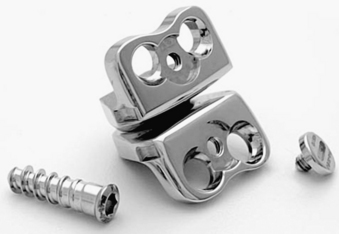
FIGURE 160-1 Prestige ST artificial cervical disc.
(Courtesy of Medtronic Sofamor Danek, Memphis, TN.)
The Prestige LP is the latest version of the Prestige artificial discs and differs from previous models in that fixation to the vertebral bodies is achieved by a set of rails that are imbedded in the vertebral end plates (Fig. 160-2). It is manufactured from a titanium-ceramic composite material that is highly durable and produces little artifact on CT and MRI. The ball-and-trough design is identical to that of the Prestige ST. A porous titanium plasma spray coating on the end-plate surface facilitates bone ingrowth and long-term fixation.
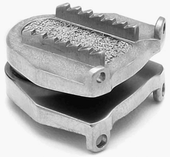
FIGURE 160-2 Prestige LP artificial cervical disc.
(Courtesy of Medtronic Sofamor Danek, Memphis, TN.)
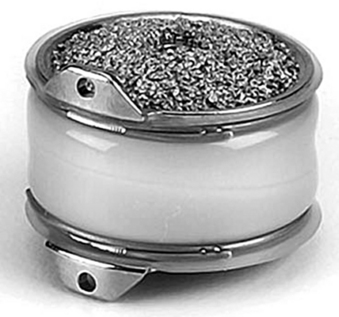
The Bryan cervical disc is designed to allow for motion similar to the normal cervical spine and resembles an actual disc (Fig. 160-3). It consists of two titanium alloy shells with a polyurethane nucleus. The bone-implant interface of each shell has an applied porous coating to facilitate ingrowth of bone and promote long-term stability. The nucleus is surrounded with a polyurethane sheath to establish a closed articulation. Sterile saline is injected into this sheath and functions as an initial lubricant. Titanium alloy seal plugs allow for retention of the saline lubricant. Small ventral flanges on the shells serve as handles to grasp the device for insertion.
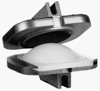
The ProDisc-C cervical disc is a metal-on-polyethylene articulating device (Fig. 160-4). This modular implant consists of two cobalt-chromium-molybdenum end plates and an ultra-high molecular weight polyethylene inlay that is preassembled into the inferior. The end plates of the prosthesis are initially secured to the vertebral bodies with central keels and have a plasma-sprayed titanium coating for long-term fixation stability.
Indications
Patients with cervical radiculopathy secondary to degenerative disc disease at one level with normal cervical spinal alignment and disc height who have failed conservative therapeutics are potential candidates for cervical arthroplasty.15 Factors to consider when selecting suitable candidates for arthroplasty include the disc height at the level of degeneration, inherent range of motion, and cervical alignment. Arthroplasty requires the dorsal elements to be intact and functional, so patients with suspected ligamentous or facet disease are not suitable candidates. Those with radiographic instability should be treated with an arthrodesis. Those with cervical kyphosis, cervical spondylolisthesis, severe multilevel spondylosis, previous laminectomy, osteoporosis, metabolic bone disease, or cervical trauma are not ideal either.16 Arthroplasty is contraindicated in the setting of significant segmental or global deformity. A recent history of infection or osteomyelitis precludes the use of a prosthetic disc device. Other relative contraindications include rheumatoid arthritis, renal failure, cancer, and chronic corticosteroid use. Multilevel cervical disc arthroplasty has not yet been evaluated prospectively.
Clinical Outcomes
There are currently seven artificial discs in the United States undergoing FDA IDE trials: Prestige ST, Prestige LP, Bryan, ProDisc-C, PCM (Cervitech, Inc., Rockaway, NJ), Kineflex (Spinal Motion, Inc., Mountain View, CA), and CerviCore (Stryker Spine, Allendale, NJ). To date, European and Australian clinical trials have demonstrated improved clinical outcomes with cervical arthroplasty compared with ACDF when using validated outcome measures such as the Neck Disability Index, an arm pain intensity visual analogue scale, and neurologic status.15,17–19 No reports of excessive wear debris, material fatigue, fracture, or failure have been reported in any of these trials.19–23 In the United States, published clinical data are available for the Prestige ST, Bryan, and ProDisc-C artificial discs.


