Capsulorrhexis and hydrodissection

Published on 08/03/2015 by admin

Filed under Opthalmology

Last modified 08/03/2015

Print this page

rate 1 star rate 2 star rate 3 star rate 4 star rate 5 star
Your rating: none, Average: 0 (0 votes)

This article have been viewed 1516 times

CHAPTER 12 Capsulorrhexis and hydrodissection

See Video image

Capsulorrhexis

Instruments

The choice of instruments for performing capsulorrhexis comprises either a bent needle referred to as a ‘cystotome’ (as was originally described) or, alternatively, capsulorrhexis forceps. A more recent addition to the choice of instruments is the femtosecond laser, at significantly greater cost compared with a bent needle.

A custom-shaped or preformed cystotome remains very popular today with many surgeons. Most use a 25 G needle. The advantages of using a needle (apart from cost) come from its small size: it can be used through a side port and causes minimal wound gape or chamber collapse. Its only disadvantage is the requirement to press downwards onto the capsule in order to gain any traction. This reduces the degrees of freedom for directional control of the tear.

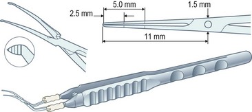
Capsulorrhexis forceps, on the other hand, allow you to apply traction in any chosen direction but are relatively bulky (particularly the early parallel-action Utratta designs) and are more likely to cause wound gape and chamber collapse than a needle. However the newer designs are greatly refined and include hinged ‘cross-action’ and coaxial designs that cause minimal wound gape and give exquisite control of the tear through incisions down to sub-2 mm (Fig. 12.1). The high outlay cost, difficulty in cleaning, and susceptibility to damage of capsulorrhexis forceps all ensure that the bent needle is here to stay.

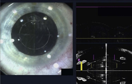
The role of lasers in surgery is assuming an ever-increasing importance and, at the time of writing, the femtosecond laser has recently been used successfully on human eyes to create high precision incisions and accurate capsulorrhexis (Fig. 12.2).

image

Fig. 12.2 Femto laser caspulorrhexis, Zoltan Nagy.

Courtesy LenSx Lasers, Inc [Aliso Viejo, CA].

In practice your choice of instrument is one of personal preference. However, it behoves all surgeons to develop the capacity for adaptability which is, after all, the basis for evolution and survival. To completely master the art of capsulorrhexis a surgeon needs to be able to use both a needle and forceps with equal facility as neither of them alone is adequate for all situations. In addition, if you can use either hand and perform the tear clockwise and anti-clockwise then no capsulorrhexis is ever going to defeat you.

Capsulorrhexis technique

Like every step in phaco the devil is in the detail and this is particularly true when performing capsulorrhexis. Careful attention is needed to the finer points of the technique because small errors at this stage can result in significant complications later on through a domino or cascade effect.

As a beginner you should start with easy eyes: the ideal scenario is a shallow orbit, well-exposed globe, good akinesia and anesthesia, clear cornea, deep chamber, well-dilated pupil, and good red reflex. If you can tick all these boxes then you are off to a great start.

Avoid being in too much of a rush to get on with the surgery. You need to ensure that the patient is comfortable and well-positioned to facilitate good coaxial illumination. Most surgeons are pleasantly surprised by how dramatically a little methylcellulose on the cornea improves visibility and it needs only an occasional top-up during surgery.

The incisions are next. I would encourage you routinely to make two side ports, one either side of the main incision, separated by about 120°. This allows you universal access from either side with a cystotome, which is very helpful, and is necessary in any case if you use bimanual I/A.

If you favor a superior approach then ensure that the speculum is rotated such that the upper blade is positioned to either straddle the tunnel (if it is an open wire style) or be on one side or the other. This avoids snagging the forceps or needle on the speculum once your instrument is inside the eye. The stage is now set.

Buy Membership for Opthalmology Category to continue reading. Learn more here