Brain stem

Published on 18/03/2015 by admin

Filed under Basic Science

Last modified 18/03/2015

Print this page

rate 1 star rate 2 star rate 3 star rate 4 star rate 5 star
Your rating: none, Average: 0 (0 votes)

This article have been viewed 7109 times

CHAPTER 19 Brain stem

The brain stem consists of the medulla oblongata, pons and midbrain. It is situated in the posterior cranial fossa, and its ventral surface lies on the clivus. It contains numerous intrinsic neuronal cell bodies and their processes, some of which are the brain stem homologues of spinal cell groups. Some brain stem cell groups are the nuclei of cranial nerves III–XII: they are concerned with the sensory, motor and autonomic innervation of the head and neck. Other autonomic fibres that arise from the brain stem are distributed more widely via the vagus nerve. The brain stem also contains a complex and sometimes ill-defined network of neurones, the reticular formation, that extends throughout its length, and is continuous caudally with its spinal counterpart. Some reticular nuclei are referred to as vital centres since they are concerned with regulation of cardiac and respiratory activities; other parts of the reticular formation are essential for cerebral cortical arousal and the maintenance of consciousness, or are involved in the regulation of muscle tone, posture and reflex activities. The brain stem is the site of termination of numerous ascending and descending fibres and is traversed by many others. The spinothalamic tract (spinal lemniscus), medial lemniscus (see Fig. 18.10) and the trigeminothalamic tracts (see Fig. 19.16) all ascend through the brain stem to reach the thalamus Prominent corticospinal projections descend through the brain stem (see Fig. 18.17) and corticobulbar projections end within it.

Damage to the brain stem is often devastating and life-threatening. This is because it is a structurally and functionally compact region, where even small lesions can destroy vital cardiac and respiratory centres, disconnect forebrain motor areas from brain stem and spinal motor neurones, and sever incoming sensory fibres from higher centres of consciousness, perception and cognition. Irreversible cardiac and respiratory arrest follow complete destruction of the neural respiratory and cardiac centres in the medulla. Clinically, this is called brain stem death, a condition that requires accurate diagnosis since it may occur in patients on life-support machines whose respiratory and cardiac functions can be artificially maintained indefinitely.

OVERVIEW OF CRANIAL NERVES AND CRANIAL NERVE NUCLEI

The cranial nerves are the routes by which the brain receives information directly from, and controls the functions of, structures which are located mainly, although not exclusively, within the head and neck. All but two of the twelve pairs of cranial nerves attach to the brain stem. Below is a brief overview of the cranial nerves (consult also Table 15.1), their associated cranial nerve nuclei, and some of the brain stem reflexes to which they contribute. The cranial nerves are described in detail on a regional basis in appropriate chapters.

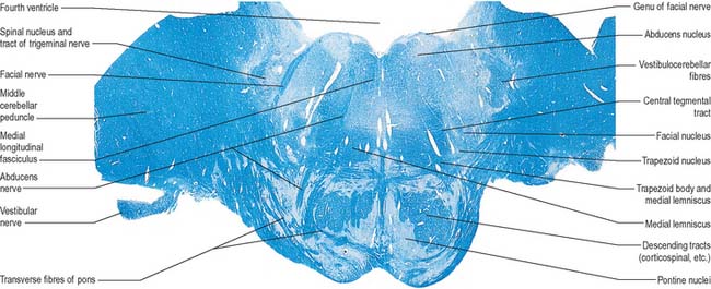
The cranial nerves are individually named and numbered (using Roman numerals) in a rostro-caudal sequence, reflecting their order of attachment to the brain. The first cranial nerve (olfactory) terminates directly in cortical and subcortical areas of the frontal and temporal lobes. It is closely associated functionally with the limbic system and is described in that context (Ch. 23). The fibres of the second cranial nerve (optic) pass into the optic chiasma where the centrally located fibres decussate; all of the fibres emerge as the optic tract, which terminates in the lateral geniculate nucleus of the thalamus. Cranial nerves III (oculomotor) and IV (trochlear) attach to the midbrain. Cranial nerve V (trigeminal) attaches to the pons, medial to the middle cerebellar peduncle. Cranial nerves VI (abducens), VII (facial) and VIII (vestibulocochlear) attach to the brain stem at, or close to, the junction of the pons with the medulla. Cranial nerves IX (glossopharyngeal), X (vagus), the cranial part of XI (accessory) and XII (hypoglossal) all attach to the medulla.

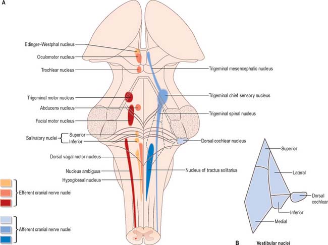
Cranial nerves III–XII, which attach to the brain stem, are associated with the brain stem cell groupings referred to collectively as the cranial nerve nuclei (Fig. 19.1). The nuclei are either the origin of efferent cranial nerve fibres or the site of termination of cranial nerve afferents. They are considered to be organized into six discontinuous longitudinal cell columns that correspond to columns that may be identified in the embryo (see Fig. 15.3). Three columns are ‘sensory’ and three are ‘motor’ in function.

The trigeminal sensory nucleus, which extends throughout the length of the brain stem and into the cervical spinal cord, represents a general somatic afferent cell column. Its principal afferents are carried in the trigeminal nerve. General visceral afferents carried by the facial, glossopharyngeal and vagus nerves end in the nucleus solitarius of the medulla. The special somatic afferent column corresponds to the vestibular and cochlear nuclei, which are located beneath the vestibular area of the floor of the fourth ventricle.

The general somatic efferent cell column consists of four nuclei that lie near the midline and give rise to motor fibres which run in nerves of the same name. From rostral to caudal, these are the oculomotor, trochlear and abducens nuclei which innervate the extraocular muscles, and the hypoglossal nucleus, which innervates all but one of the muscles of the tongue. The general visceral efferent, or parasympathetic, cell column is made up of the Edinger–Westphal nucleus of the midbrain, salivary nuclei of the pons, and vagal nucleus of the medulla. Cells in the special visceral efferent column innervate muscles derived from the branchial arches, and lie in the trigeminal motor nucleus, the facial nucleus and the nucleus ambiguus.

MEDULLA OBLONGATA

EXTERNAL FEATURES AND RELATIONS

The medulla oblongata extends from just above the first pair of cervical spinal nerves to the lower border of the pons (Fig. 28.11). It is approximately 3 cm in length and 2 cm in diameter at its widest. The ventral surface of the medulla is separated from the basilar part of the occipital bone and apex of the dens by the meninges and occipito-axial ligaments. Caudally, the dorsal surface of the medulla occupies the midline notch between the cerebellar hemispheres.

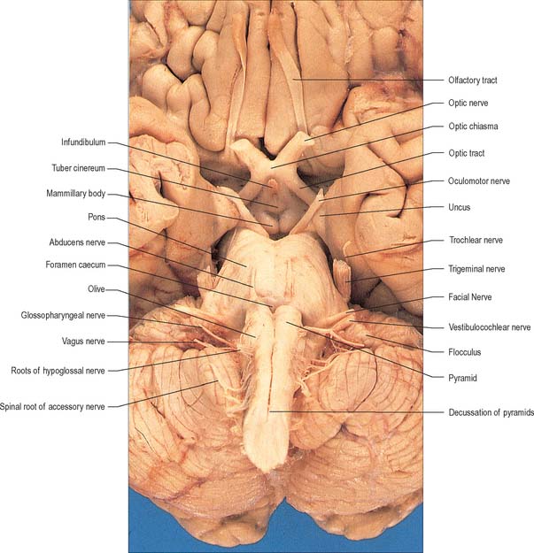
The ventral and dorsal surfaces of the medulla (Fig. 19.2, Fig. 19.3) possess a longitudinal median fissure and sulcus, respectively, which are continuous with their spinal counterparts. Caudally, the ventral median fissure is interrupted by the obliquely crossing fascicles of the pyramidal decussation. Rostrally, it ends at the pontine border in a diminutive depression, the foramen caecum. Immediately lateral to the ventral median fissure there is a prominent elongated ridge, the pyramid, which contains descending pyramidal, or corticospinal, axons. The lateral margin of the pyramid is indicated by a shallow ventrolateral sulcus. From this emerges, in line with the ventral spinal nerve roots, a linear series of rootlets which constitute the hypoglossal nerve. The abducens nerve emerges at the slightly narrowed rostral end of the pyramid, where it adjoins the pons. Caudally the pyramid tapers into the spinal ventral funiculus. Lateral to the pyramid and the ventrolateral sulcus there is an oval prominence, the olive (Fig. 19.2), which contains the inferior olivary nucleus. Lateral to the olive is the posterolateral sulcus. The glossopharyngeal, vagus and accessory nerves join the brain stem along the line of this sulcus, in line with the dorsal spinal nerve roots.

The spinal central canal extends into the caudal half of the medulla, migrating progressively more dorsally until it opens out into the lumen of the fourth ventricle. This divides the medulla into a closed part, which contains the central canal, and an open part, which contains the caudal half of the fourth ventricle (Fig. 19.3).

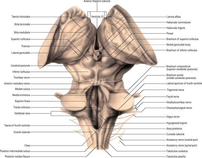
In the closed part of the medulla, a shallow dorsal intermediate sulcus, on either side of the dorsal median sulcus and continuous with its cervical spinal counterpart, indicates the location of the ascending dorsal columns (fasciculus gracilis and fasciculus cuneatus). The ascending fasciculi are at first parallel to each other, but at the caudal end of the fourth ventricle they diverge, and each develops an elongated swelling, the gracile and cuneate tubercles, produced by the subjacent nuclei gracilis and cuneatus respectively (Fig. 19.3, Fig. 19.4, Fig. 19.5). Most fibres in the fasciculi synapse with neurones in their respective nuclei, and these project to the contralateral thalamus, which, in turn, projects to the primary somaesthetic cortex (see Fig. 18.10). The inferior cerebellar peduncle forms a rounded ridge between the caudal part of the fourth ventricle and the glossopharyngeal and vagal rootlets. The peduncles of the two sides diverge and incline to enter the cerebellar hemispheres, where they are crossed by the striae medullares which run to the dorsal median sulcus of the ventricular floor (Fig. 19.3). Here also the peduncles form the anterior and rostral boundaries of the lateral recess of the fourth ventricle. This becomes continuous with the subarachnoid space through the lateral apertures of the fourth ventricle, or foramina of Luschka. A tuft of choroid plexus, continuous with that of the fourth ventricle, protrudes from the foramina on either side.

INTERNAL STRUCTURE

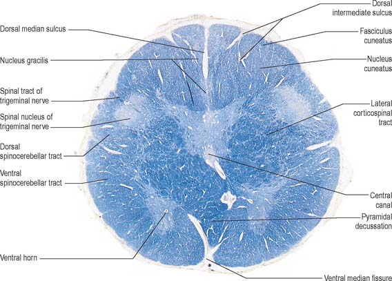
Transverse section of the medulla at the level of the pyramidal decussation

A transverse section across the lower medulla oblongata (Fig. 19.4) intersects the dorsal, lateral and ventral funiculi, which are continuous with their counterparts in the spinal cord. The ventral funiculi are separated from the central grey matter by corticospinal fibres, which cross in the pyramidal decussation to reach the contralateral lateral funiculi (see Fig. 19.8). The decussation displaces the central grey matter and central canal dorsally. Continuity between the ventral grey column and central grey matter, which is maintained throughout the spinal cord, is lost. The column subdivides into the supraspinal nucleus (continuous above with that of the hypoglossal nerve), which is the efferent source of the first cervical nerve, and the spinal nucleus of the accessory nerve, which provides some spinal accessory fibres and merges rostrally with the nucleus ambiguus.

The dorsal grey column is also modified at this level as the nucleus gracilis appears as a grey lamina in the ventral part of the fasciculus gracilis. The nucleus begins caudal to the nucleus cuneatus, which invades the fasciculus cuneatus from its ventral aspect in similar fashion.

The spinal nucleus and spinal tract of the trigeminal nerve are visible ventrolateral to the dorsal columns. They are continuous with the substantia gelatinosa and tract of Lissauer of the spinal cord.

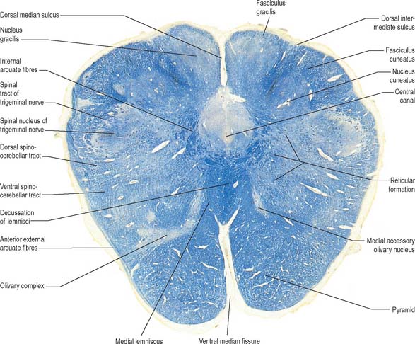
Transverse section of the medulla at the level of the decussation of the medial lemniscus

The medullary white matter is rearranged above the level of the pyramidal decussation (Fig. 19.5). The pyramids form two large ventral bundles flanking the ventral median fissure on the ventral surface of the medulla.

They contain corticospinal fibres of ipsilateral origin. The nucleus gracilis is prominent on the dorsal aspect, with diminishing numbers of fibres of the fasciculus gracilis located on its dorsal, medial and lateral margins. The nucleus cuneatus is well developed. Both nuclei retain continuity with the central grey matter at this level, but this is lost more rostrally. First-order afferent fibres contained within the fasciculi gracilis and cuneatus synapse upon neurones in their respective nuclei. Second-order axons emerge from the nuclei as internal arcuate fibres, at first curving ventrolaterally around the central grey matter and then ventromedially between the spinal tract of the trigeminal nerve and the central grey matter. The fibres decussate in the midline thereafter forming the medial lemniscus which ascends to the thalamus. The decussation of internal arcuate fibres is located dorsal to the pyramids and ventral to the central grey matter, which is therefore more dorsally displaced than in the previous section.

The medial lemniscus ascends from the decussation as a flattened tract, near the median raphe. The pyramidal tract lies ventrally, and the medial longitudinal fasciculus and the tectospinal tract lie dorsally. Decussating lemniscal fibres are rearranged as they cross, so that those derived from the nucleus gracilis come to lie ventral to those from the nucleus cuneatus. Above this level, the medial lemniscus is further rearranged so that ventral (gracile) fibres migrate laterally, whilst dorsal (cuneate) fibres migrate medially. After such rearrangement, the medial lemniscus is somatotopically organized with C1 to S4 spinal segments represented sequentially from medial to lateral.

The nucleus of the spinal tract of the trigeminal nerve is separated from the central grey matter by internal arcuate fibres, and from the lateral medullary surface by the spinal tract of the trigeminal and by some dorsal spinocerebellar tract fibres. The latter progressively incline dorsally, and enter the inferior cerebellar peduncle at a higher level.

Transverse section of the medulla at the caudal end of the fourth ventricle

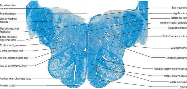
A transverse section at the lower end of the fourth ventricle (Fig. 19.6) shows some new features together with most of those already described. The total area of grey matter is increased by the presence of the large olivary nuclear complex and nuclei of the vestibulocochlear, glossopharyngeal, vagus and accessory nerves.
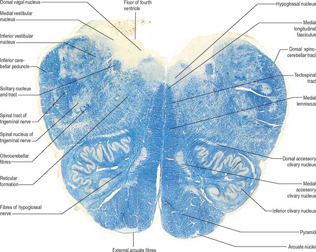
A smooth, oval elevation, the olive, lies between the ventrolateral and dorsolateral sulci of the medulla. It is formed by the underlying inferior olivary complex of nuclei, and lies lateral to the pyramid, separated from it by the ventrolateral sulcus and emerging hypoglossal nerve fibres. The roots of the facial nerve emerge between its rostral end and the lower pontine border, in the cerebellopontine angle. The arcuate nuclei are curved, interrupted bands, ventral to the pyramids, and are said to be displaced pontine nuclei. Anterior external arcuate fibres and those of the striae medullares are derived from them. They project mainly to the contralateral cerebellum through the inferior cerebellar peduncle (Fig. 19.7).

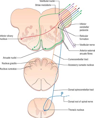
The inferior olivary nucleus is an irregularly crenated mass of grey matter with a medially directed hilum, through which numerous fibres enter and leave the nucleus. It has prominent connections with the cerebellum and is described more fully in Chapter 20.

The central grey matter at this level constitutes the ventricular floor. It contains (sequentially from medial to lateral): the hypoglossal nucleus, dorsal motor nucleus of the vagus, nucleus solitarius, and the caudal ends of the inferior and medial vestibular nuclei.

The tractus solitarius and the associated nucleus solitarius extend throughout the length of the medulla. The tract is composed of general visceral afferents from the vagus and glossopharyngeal nerves. The nucleus and its central connections with the reticular formation subserve the reflex control of cardiovascular, respiratory and cardiac functions. The rostral portion of the tract consists of gustatory fibres from the facial, glossopharyngeal and vagal nerves; they project to the rostral pole of the nucleus solitarius, which is sometimes referred to as the gustatory nucleus.

The medial longitudinal fasciculus is a small compact tract near the midline, ventral to the hypoglossal nucleus, that is continuous with the ventral vestibulospinal tract. At this medullary level it is displaced dorsally by the pyramidal and lemniscal decussations. It ascends in the pons and midbrain, maintaining its relationship to the central grey matter and midline, and is therefore near the somatic efferent nuclear column. Fibres from a variety of sources course for short distances in the tract.

The spinocerebellar, spinotectal, vestibulospinal, rubrospinal and lateral spinothalamic (spinal lemniscal) tracts all lie in the ventrolateral area of the medulla at this level. The tracts are limited dorsally by the nucleus of the spinal tract of the trigeminal and ventrally by the pyramid.
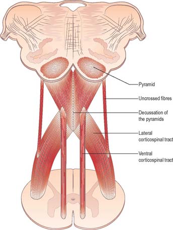
Numerous islets of grey matter are scattered centrally in the ventrolateral medulla, an area intersected by nerve fibres that run in all directions. This is the reticular formation, which exists throughout the medulla and extends into the pontine tegmentum and midbrain.

Pyramidal tract

Each pyramid contains descending corticospinal fibres, derived from the ipsilateral cerebral cortex, which have traversed the internal capsule, midbrain and pons (Fig. 19.8). Approximately 70–90% of the axons leave the pyramids in successive bundles, crossing in and deep to the ventral median fissure as the pyramidal decussation. In the rostral medulla fibres cross by inclining ventromedially, whereas more caudally they pass dorsally, decussating ventral to the central grey matter. The decussation is orderly, such that fibres destined to end in the cervical segments cross first. Fibres continue to pass dorsally as they descend, and reach the contralateral spinal lateral funiculus as the crossed lateral corticospinal tract. Most uncrossed corticospinal fibres descend ventromedially in the ipsilateral ventral funiculus, as the ventral corticospinal tract. A minority run dorsolaterally to join the lateral corticospinal tracts as a small uncrossed component. The corticospinal tracts display somatotopy at almost all levels. In the pyramids the arrangement is like that at higher levels, in that the most lateral fibres subserve the most medial arm and neck movements. Similar somatotopy is ascribed to the lateral corticospinal tracts within the spinal cord.

Dorsal column nuclei

The nuclei gracilis and cuneatus are part of the pathway that is the major route for discriminative aspects of tactile and proprioceptive sensation. The upper regions of both nuclei are reticular and contain small and large multipolar neurones with long dendrites. The lower regions contain clusters of large round neurones with short and profusely branching dendrites. Upper and lower zones differ in their connections but both receive terminals from the dorsal spinal roots at all levels. Dorsal funicular fibres from neurones in the spinal grey matter terminate only in the superior, reticular zone. Variable ordering and overlap of terminals, on the basis of spinal root levels, occur in both zones. The lower extremity is represented medially, the trunk ventrally, and the digits dorsally. There is modal specificity, i.e. lower levels respond to low-threshold cutaneous stimuli, and upper reticular levels to inputs from fibres serving receptors in the skin, joints and muscles. The cuneate nucleus is divided into several parts. Its middle zone contains a large pars rotunda, in which rostrocaudally elongated medium-sized neurones are clustered between bundles of densely myelinated fibres. The reticular poles of its rostral and caudal zones contain scattered, but evenly distributed, neurones of various sizes. The pars triangularis is smaller and laterally placed.

There is a somatotopic pattern of termination of cutaneous inputs from the upper limb upon the cell clusters of the pars rotunda. Terminations are diffuse in the reticular poles.

The gracile and cuneate nuclei serve as relays between the spinal cord and higher levels. Primary spinal afferents synapse with multipolar neurones in the nuclei that form the major nuclear efferent projection. The nuclei also contain interneurones, many of which are inhibitory. Descending afferents from the somatosensory cortex reach the nuclei through the corticobulbar tracts, and appear to be restricted to the upper, reticular zones. These afferents both inhibit and enhance activity and are believed to be involved in sensory modulation. The reticular zones also receive connections from the reticular formation.

Neurones of dorsal column nuclei receive terminals of long, uncrossed, primary afferent fibres of the fasciculi gracilis and cuneatus, which carry information concerning deformation of skin, movement of hairs, joint movement and vibration. Unit recording of the neurones in dorsal column nuclei shows that their tactile receptive fields (i.e. the skin area in which a response can be elicited) vary in size, although they are mostly small, and are smallest for the digits. Some fields have excitatory centres and inhibitory surrounds, which means that stimulation just outside its excitatory field inhibits the neurone. Neurones in the nuclei are spatially organized into a somatotopic map of the periphery (in accord with the similar localization in the dorsal columns). In general, specificity is high. Many cells receive input from one or a few specific receptor types, e.g. hair, type I and II slowly adapting receptors and Pacinian corpuscles, and some cells respond to Ia muscle spindle input. However, some neurones receive convergent input from tactile pressure and hair follicle receptors.

A variety of control mechanisms can modulate the transmission of impulses through the dorsal column–medial lemniscus pathway. Concomitant activity in adjacent dorsal column fibres may result in presynaptic inhibition by depolarization of the presynaptic terminals of one of them. Stimulation of the sensory–motor cortex also modulates the transmission of impulses by both pre- and postsynaptic inhibitory mechanisms, and sometimes by facilitation. These descending influences are mediated by the corticospinal tract. Modulation of transmission by inhibition also results from stimulation of the reticular formation, raphe nuclei and other sites.

The accessory cuneate nucleus, dorsolateral to the cuneate nucleus, is part of the spinocerebellar system of precerebellar nuclei (Fig. 19.7). It contains large neurones like those in the spinal thoracic nucleus and receives the lateral fibres of the fasciculus cuneatus, which carry proprioceptive impulses from the upper limb that enter the cervical spinal cord rostral to the thoracic nucleus. The accessory cuneate neurones give rise to the posterior external arcuate fibres that enter the cerebellum by the ipsilateral inferior cerebellar peduncle in the cuneocerebellar tract. A group of neurones, nucleus Z, identified in animals between the upper pole of the nucleus gracilis and the inferior vestibular nucleus, is said to be present in the human medulla. Its input is probably from the dorsal spinocerebellar tract, which carries proprioceptive information from the ipsilateral lower limb, and it projects through internal arcuate fibres to the contralateral medial lemniscus.

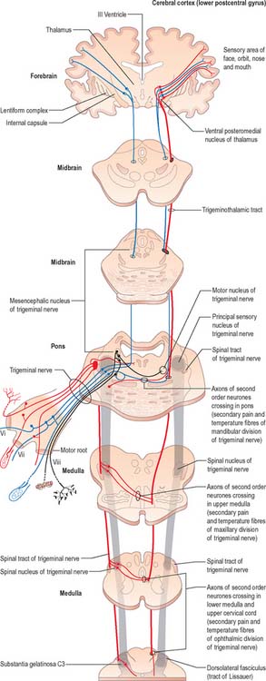
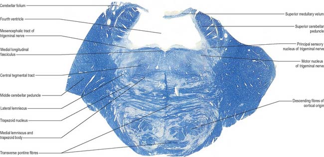
Trigeminal sensory nucleus

The trigeminal sensory nucleus receives the primary afferents of the trigeminal nerve. It is a large nucleus, and extends caudally into the cervical spinal cord and rostrally into the midbrain. The principal and largest division of the nucleus is located in the pontine tegmentum.

On entering the pons, the fibres of the sensory root of the trigeminal nerve run dorsomedially towards the principal sensory nucleus, which is situated at this level (Fig. 19.9). Before reaching the nucleus approximately 50% of the fibres divide into ascending and descending branches – the others ascend or descend without division. The descending fibres, of which 90% are less than 4 μm in diameter, form the spinal tract of the trigeminal nerve, which reaches the upper cervical spinal cord. The tract embraces the nucleus of the spinal tract of the trigeminal (spinal trigeminal nucleus; Fig. 19.4, Fig. 19.5, Fig. 19.6, Fig. 19.10, Fig. 19.11). There is a precise somatotopic organization in the tract. Fibres from the ophthalmic division of the trigeminal lie ventrolaterally, those from the mandibular division lie dorsomedially, and the maxillary fibres lie between them. The tract is completed on its dorsal rim by fibres from the sensory roots of the facial, glossopharyngeal and vagus nerves. All of these fibres synapse in the nucleus caudalis.

The detailed anatomy of the spinal tract of the trigeminal excited early clinical interest because it was recognized that dissociated sensory loss could occur in the trigeminal area. For example, in Wallenberg’s syndrome, occlusion of the posterior inferior cerebellar branch of the vertebral artery leads to loss of pain and temperature sensation in the ipsilateral half of the face with retention of common sensation. Neurosurgery in this region, as early as the 1890s, attempted to alleviate paroxysmal trigeminal neuralgia. The introduction of medullary tractotomy confirmed that dissociated thermoanalgesia of the face was associated with destruction of the tract.

There are conflicting opinions on the pattern of termination of the fibres in the spinal nucleus. It has long been held that fibres are organized rostrocaudally within the tract. According to this view, ophthalmic fibres are ventral and descend to the lower limit of the first cervical spinal segment, maxillary fibres are central and do not extend below the medulla oblongata, whilst mandibular fibres are dorsal and do not extend much below the mid-medullary level. The results of section of the spinal tract in cases of severe trigeminal neuralgia support this distribution. It was found that a section 4 mm below the obex rendered the ophthalmic and maxillary areas analgesic but tactile sensibility, apart from the abolition of ‘tickle’, was much less affected. To include the mandibular area it was necessary to section at the level of the obex. More recently, it has been proposed that fibres are arranged dorsoventrally within the spinal tract. There appear to be sound anatomico-physiological and clinical reasons for believing that all divisions terminate throughout the whole nucleus, although the ophthalmic division may not project fibres as far caudally as the maxillary and mandibular divisions. Fibres from the posterior face (adjacent to C2) terminate in the lower (caudal) part, whilst those from the upper lip, mouth and nasal tip terminate at a higher level. This can give rise to a segmental (cross-divisional) sensory loss in syringobulbia. Tractotomy of the spinal tract, if carried out at a lower level, can spare the perioral region, a finding that would accord with the ‘onion-skin’ pattern of loss of pain sensation. However, in clinical practice, the progression of anaesthesia on the face is most commonly ‘divisional’ rather than onion-skin in distribution.

Fibres of the glossopharyngeal, vagus and facial nerves subserving common sensation (general visceral afferent) form a column dorsally within the spinal tract of the trigeminal nerve and synapse with cells in the lowest part of the spinal trigeminal nucleus. Consequently, operative section of the dorsal part of the spinal tract results in analgesia that extends to the mucosa of the tonsillar sinus, the posterior third of the tongue and adjoining parts of the pharyngeal wall (glossopharyngeal nerve), and the cutaneous area supplied by the auricular branch of the vagus.

Other afferents that reach the spinal nucleus are from the dorsal roots of the upper cervical nerves and from the sensorimotor cortex.

The spinal nucleus is considered to consist of three parts: the subnucleus oralis (which is most rostral and adjoins the principal sensory nucleus); the subnucleus interpolaris; and the subnucleus caudalis (which is the most caudal part and is continuous below with the dorsal grey column of the spinal cord). The structure of the subnucleus caudalis is different from that of the other subnuclei. It has a structure analogous to that of the dorsal horn of the spinal cord, with a similar arrangement of cell laminae, and is involved in trigeminal pain perception. Cutaneous nociceptive afferents and small-diameter muscle afferents terminate in layers I, II, V and VI of the subnucleus caudalis. Low-threshold mechanosensitive afferents of Aβ neurones terminate in layers III and IV of the subnucleus caudalis and rostral (interpolaris, oralis and main sensory) nuclei.

Many of the neurones in the subnucleus caudalis that respond to cutaneous or tooth-pulp stimulation are also excited by stimulation of jaw or tongue muscles. This indicates that convergence of superficial and deep afferent inputs via wide-dynamic-range or nociceptive-specific neurones occurs in the nucleus. Similar convergence of superficial and deep inputs occurs in the rostral nuclei and may account for the poor localization of trigeminal pain, and for the spread of pain, which often makes diagnosis difficult.

There are distinct subtypes of cells in lamina II. Afferents from ‘higher-centres’ arborize within it, as do axons from nociceptive and low-threshold afferents. Descending influences from these higher centres include fibres from the periaqueductal grey matter and from the nucleus raphe magnus and associated reticular formation.

The nucleus raphe magnus projects directly to the subnucleus caudalis, probably via enkephalin, noradrenaline and 5-HT-containing terminals. These fibres directly or indirectly (through local interneurones) influence pain perception. Stimulation of periaqueductal grey matter or nucleus raphe magnus inhibits the jaw opening reflex to nociception, and may induce primary afferent depolarization in tooth-pulp afferents and other nociceptive facial afferents. Neurones in the subnucleus caudalis can be suppressed by stimuli applied outside their receptive field, particularly by noxious stimuli. The subnucleus caudalis is an important site for relay of nociceptive input and functions as part of the pain ‘gate-control’. However, rostral nuclei also have a nociceptive role. Tooth-pulp afferents via wide-dynamic-range and nociceptive-specific neurones may terminate in rostral nuclei, which all project to the subnucleus caudalis.

Most fibres arising in the trigeminal sensory nuclei cross the midline and ascend in the trigeminal lemniscus. They end in the contralateral thalamic nucleus ventralis posterior medialis, from which third-order neurones project to the cortical postcentral gyrus (areas 1, 2 and 3). However, some trigeminal nucleus efferents ascend to the nucleus ventralis posterior medialis of the ipsilateral thalamus.

Fibres from the subnucleus caudalis, especially from laminae I, V and VI, also project to the rostral trigeminal nuclei, cerebellum, periaqueductal grey of the midbrain, parabrachial area of the pons, the brain stem reticular formation and the spinal cord. Fibres from lamina I project to the subnucleus medius of the medial thalamus.

Hypoglossal nucleus

The prominent hypoglossal nucleus lies near the midline in the dorsal medullary grey matter. It is approximately 2 cm long. Its rostral part lies beneath the hypoglossal triangle in the floor of the fourth ventricle (Fig. 19.3) and its caudal part extends into the closed part of the medulla.

The hypoglossal nucleus consists of large motor neurones interspersed with myelinated fibres. It is organized into dorsal and ventral nuclear tiers, each divisible into medial and lateral subnuclei. There is a musculotopic organization of motor neurones within the nuclei that corresponds to the structural and functional divisions of tongue musculature. Thus, motor neurones innervating tongue retrusor muscles are located in dorsal/dorsolateral subnuclei, whereas motor neurones innervating the main tongue protrusor muscle are located in ventral/ventromedial regions of the nucleus. Although relatively little is known about the organization of motor neurones innervating the intrinsic muscles of the tongue, experimental evidence suggests that motor neurones of the medial division of the hypoglossal nucleus innervate tongue muscles that are oriented in planes transverse to the long axis of the tongue (transverse and vertical intrinsics and genioglossus), whereas motor neurones of the lateral division innervate tongue muscles that are oriented parallel to this axis (styloglossus, hyoglossus, superior and inferior longitudinal).

Several smaller groups of cells lie near the hypoglossal nucleus (perihypoglossal nuclei), but none is known for certainty to be connected with the hypoglossal nerve or nucleus. They include the nucleus intercalatus, sublingual nucleus, nucleus prepositus hypoglossi and nucleus paramedianus dorsalis (reticularis). Gustatory and visceral connections are attributed to the nucleus intercalatus.

Hypoglossal fibres emerge ventrally from their nucleus, traverse the reticular formation lateral to the medial lemniscus, pass medial to (sometimes through) the inferior olivary nucleus, and curve laterally to emerge as a linear series of 10–15 rootlets in the ventrolateral sulcus between the pyramid and olive.

The hypoglossal nucleus receives corticobulbar fibres from the precentral gyrus and adjacent areas of (mainly) the contralateral hemisphere. They either synapse on motor neurones of the nucleus directly or on interneurones. Evidence indicates that the most medial hypoglossal subnuclei receive projections from both hemispheres. The nucleus may connect with the cerebellum via adjacent perihypoglossal nuclei, and perhaps also with the medullary reticular formation, the trigeminal sensory nuclei and the solitary nucleus.

Inferior olivary nucleus

The olivary nuclear complex consists of the large inferior olivary nucleus and the much smaller medial accessory and dorsal accessory olivary nuclei (Fig. 19.6). They are the so-called precerebellar nuclei, a group that also includes the pontine, arcuate, vestibular, reticulocerebellar and spinocerebellar nuclei, all of which receive afferents from specific sources and project to the cerebellum. The inferior olivary nucleus contains small neurones, most of which form the olivocerebellar tract, which emerges either from the hilum or through the adjacent grey matter, to run medially and intersect the medial lemniscus (Fig. 19.6). Its fibres cross the midline, and sweep either dorsal to, or through, the opposite olivary nucleus. They intersect the lateral spinothalamic and rubrospinal tracts and the spinal trigeminal nucleus, and enter the contralateral inferior cerebellar peduncle, where they constitute its major component. Fibres from the contralateral inferior olivary complex terminate on Purkinje cells in the cerebellum as climbing fibres – there is a one-to-one relationship between Purkinje cells and neurones in the complex. Afferent connections to the inferior olivary nucleus are both ascending and descending. Ascending fibres, mainly crossed, arrive from all spinal levels in the spino-olivary tracts and via the dorsal columns. Descending ipsilateral fibres come from the cerebral cortex, thalamus, red nucleus and central grey of the midbrain. In part the two latter projections make up the central tegmental tract (fasciculus).

The medial accessory olivary nucleus is a curved grey lamina, concave laterally, between the medial lemniscus and pyramid and the ventromedial aspect of the inferior olivary nucleus. The dorsal accessory olivary nucleus is a similar lamina, dorsomedial to the inferior olivary nucleus. Both nuclei are connected to the cerebellum. The accessory olivary nuclei are phylogenetically older than the inferior olivary nucleus, and they are connected with the paleocerebellum. In all connections, cerebral, spinal and cerebellar, the olivary nuclei display specific topographical organization (Ch. 20).
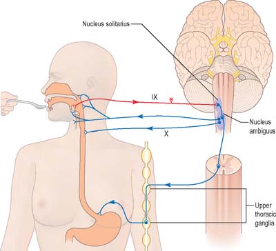
Nucleus solitarius

The nucleus solitarius (solitary nucleus, nucleus of the solitary tract) lies ventrolateral to the vagal nucleus and is almost coextensive with it. A neuronal group ventrolateral to the nucleus solitarius has been termed the nucleus parasolitarius. The nucleus solitarius is intimately related to, and receives fibres from, the tractus solitarius, which carries afferent fibres from the facial, glossopharyngeal and vagus nerves. These fibres enter the tract in descending order and convey gustatory information from the lingual and palatal mucosa. They may also convey visceral impulses from the pharynx (glossopharyngeal and vagus) and from the oesophagus and abdominal alimentary canal (vagus). There is some overlap in this vertical representation.

Termination of special visceral gustatory afferents within the nucleus shows a viscerotopic pattern, predominantly in the rostral region. Experimental evidence suggests that fibres from the anterior two-thirds of the tongue and the roof of the oral cavity (which travel via the chorda tympani and greater petrosal branches of the facial nerve) terminate in the extreme rostral part of the solitary complex. Those from the circumvallate and foliate papillae of the posterior third of the tongue, tonsils, palate and pharynx (which travel via the lingual branch of the glossopharyngeal nerve) are distributed throughout the rostrocaudal extent of the nucleus, predominantly rostral to the obex. Gustatory afferents from the larynx and epiglottis (which travel via the superior laryngeal branch of the vagus) have a more caudal and lateral distribution. The nucleus solitarius may also receive fibres from the spinal cord, cerebral cortex and cerebellum.

Medial and commissural subnuclei in the caudal part of the nucleus appear to be the primary site of termination for gastrointestinal afferents. Ventral and interstitial subnuclei probably receive tracheal, laryngeal and pulmonary afferents and play an important role in both respiratory control and possibly rhythm generation. The carotid sinus and aortic body nerves terminate in the dorsal and dorsolateral region of the nucleus solitarius, which may be involved in cardiovascular regulation.

The nucleus solitarius is thought to project to the sensory thalamus and thence to the cerebral cortex. It may also project to the upper levels of the spinal cord through a solitariospinal tract. Secondary gustatory axons cross the midline. Many subsequently ascend the brain stem in the dorsomedial part of the medial lemniscus and synapse on the most medial neurones of the thalamic nucleus ventralis posterior medialis (in a region sometimes termed the accessory arcuate nucleus). Axons from the nucleus ventralis posterior medialis radiate through the internal capsule to the anteroinferior area of the sensorimotor cortex and the insula. It is thought that other ascending paths end in a number of the hypothalamic nuclei, and so mediate the route by which gustatory information may reach the limbic system and allow appropriate autonomic reactions to be made.

Swallowing and gag reflexes

During the normal processes of eating and drinking, passage of material to the rear of the mouth stimulates branches of the glossopharyngeal nerve in the oropharynx (Fig. 19.12). This information is relayed via the nucleus solitarius to the nucleus ambiguus, which contains the motor neurones innervating the muscles of the palate, pharynx, and larynx. The nasopharynx is closed off from the oropharynx by elevation of the soft palate. The larynx is raised, its entrance narrowed and the glottis is closed. Peristaltic activity down the oesophagus to the stomach is mediated through the pharyngeal plexus.

If stimulation of the oropharynx occurs, other than during swallowing, the gag reflex may be initiated. There is a reflex contraction of the muscles of the pharynx, soft palate, and fauces that, if extreme, may result in retching and vomiting.

Nucleus ambiguus

The nucleus ambiguus is a group of large motor neurones, situated deep in the medullary reticular formation (see Fig. 19.10). It extends rostrally as far as the upper end of the vagal nucleus while caudally it is continuous with the nucleus of the spinal accessory nerve. Fibres emerging from it pass dorsomedially, then curve laterally. Rostral fibres join the glossopharyngeal nerve. Caudal fibres join the vagus and cranial accessory nerves and are distributed to the pharyngeal constrictors, intrinsic laryngeal muscles and striated muscles of the palate and upper oesophagus.

The nucleus ambiguus contains several cellular subgroups, and some topographical representation of the muscles innervated has been established. Individual laryngeal muscles are innervated by relatively discrete groups of cells in more caudal zones. Neurones that innervate the pharynx lie in the intermediate area, and neurones that innervate the oesophagus and soft palate are rostral.

The nucleus ambiguus receives corticobulbar fibres bilaterally and is connected to many brain stem centres. At its upper end, a small retrofacial nucleus intervenes between it and the facial nucleus. Although the nucleus ambiguus is generally regarded as a special visceral efferent nucleus, it is also a reputed source of general visceral efferent fibres to the vagus.